Researchers in Hungary have taken a fresh look at ancient Egyptian mummified remains using a new generation of CT imaging, producing a level of detail not achieved in earlier scans. The work focuses on specimens held by the Semmelweis Museum of Medical History, part of the Hungarian National Museum Public Collection Center. These remains, some more than 2,300 years old, include two heads, two left lower limbs, a hand, and a bundle once thought to contain a different type of object.
The scans were carried out at Semmelweis University’s Medical Imaging Center with a CT system equipped with a photon-counting detector. The examinations took place at night, outside normal clinical hours. This technology records X-ray signals in a more precise way than conventional systems, which helps separate fine structural details within layered materials such as wrappings, bone, and soft tissue residues.
Earlier imaging campaigns had already examined parts of this collection, though older equipment limited the level of detail. Radiocarbon dating had confirmed that at least some of the remains date to between 401 and 259 BCE. The new scans revisit these same objects with sharper resolution, allowing researchers to reassess earlier interpretations.
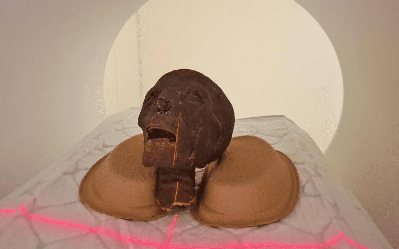
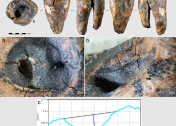
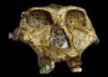
The internal structure of the mummified heads has now been mapped with greater clarity, especially in the teeth and skull sutures. These features help estimate age at death. The improved data also support future digital reconstructions of the skulls, with the possibility of facial reconstructions based on anatomical evidence.
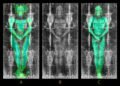
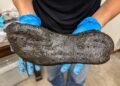
The lower limbs offered new clues as well. One specimen, previously difficult to diagnose, now shows signs consistent with osteoporosis. Researchers are working to determine whether this condition developed with age or resulted from disease. A second limb appears to belong to a younger individual, though a precise age range has not yet been established.


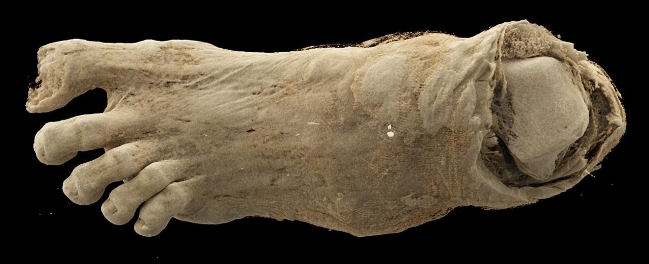
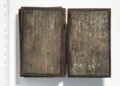
One of the more surprising findings concerns a wrapped bundle that had been misidentified in the past. Initial visual inspection suggested a human head, and later a bird mummy. Earlier CT imaging corrected this to a human foot. The latest scans go further, revealing multiple layers of bandaging with distinct structural patterns. These details provide information about embalming practices and the handling of the body after death. The reason why this foot became separated from the rest of the body remains unclear.
The mummified hand included in the study also draws attention. Researchers are examining bone size and development to determine whether it belonged to a child or an adult. The same data could help estimate sex and age.
Work on the dataset continues, with detailed evaluation still in progress. The team expects that the high-resolution images will support more accurate diagnoses and a better understanding of both health conditions and mummification techniques in ancient Egypt.
More information: Semmelweis University


Disclaimer: This website is a science-focused magazine that welcomes both academic and non-academic audiences. Comments are written by users and may include personal opinions or unverified claims. They do not necessarily reflect the views of our editorial team or rely on scientific evidence.
Comment Policy: We kindly ask all commenters to engage respectfully. Comments that contain offensive, insulting, degrading, discriminatory, or racist content will be automatically removed.